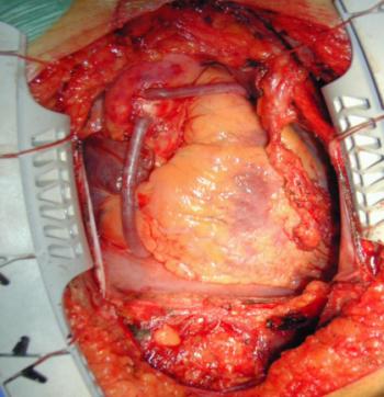
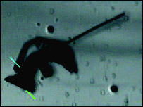
Heart valve surgery – operation for replacement heart valves
[media id=83 width=500 height=400]
heart valves maintain the unidirectional flow of blood in the heart by opening and closing depending on the difference in pressure on each side. They are mechanically similar to reed valves.
Atrioventricular valves
These are small valves that prevent backflow from the ventricles into the atria during systole. They are anchored to the wall of the ventricle by chordae tendineae, which prevent the valve from inverting.
The chordae tendineae are attached to papillary muscles that cause tension to better hold the valve. Together, the papillary muscles and the chordae tendineae are known as the subvalvular apparatus. The function of the subvalvular apparatus is to keep the valves from prolapsing into the atria when they close. The subvalvular apparatus have no effect on the opening and closure of the valves, however. This is caused entirely by the pressure gradient across the valve.
The closure of the AV valves is heard as the first heart sound
Echocardiography — The echocardiogram is an ultrasound of the heart. Using standard ultrasound techniques, two-dimensional slices of the heart can be imaged.
Vein — In biology, a vein is a blood vessel which carries blood toward the heart. Veins form part of the circulatory system
The tricuspid valve is the three flapped valve on the right side of the heart, between the right atrium and the right ventricle which stops the backflow of blood between the two. It has three cusps.
Semilunar valves
These are located at the base of both the pulmonary trunk (pulmonary artery) and the aorta, the two arteries taking blood out of the ventricles. These valves permit blood to be forced into the arteries, but prevent backflow of blood from the arteries into the ventricles. [1] These valves do not have chordae tendineae, and are more similar to valves in veins than atrioventricular valves.
Aortic valve
The aortic valve lies between the left ventricle and the aorta. The aortic valve has three cusps. During ventricular systole, pressure rises in the left ventricle. When the pressure in the left ventricle rises above the pressure in the aorta, the aortic valve opens, allowing blood to exit the left ventricle into the aorta. When ventricular systole ends, pressure in the left ventricle rapidly drops. When the pressure in the left ventricle decreases, the aortic pressure forces the aortic valve to close. The closure of the aortic valve contributes the A2 component of the second heart sound (S2).
The most common congenital abnormality of the heart is the bicuspid aortic valve. In this condition, instead of three cusps, the aortic valve has two cusps. This condition is often undiagnosed until the person develops calcific aortic stenosis. Aortic stenosis occurs in this condition usually in patients in their 40s or 50s, an average of over 10 years earlier than in people with normal aortic valves.
Pulmonary valve
The pulmonary valve (sometimes referred to as the pulmonic valve) is the semilunar valve of the heart that lies between the right ventricle and the pulmonary artery and has three cusps. Similar to the aortic valve, the pulmonary valve opens in ventricular systole, when the pressure in the right ventricle rises above the pressure in the pulmonary artery. At the end of ventricular systole, when the pressure in the right ventricle falls rapidly, the pressure in the pulmonary artery will close the pulmonary valve.
The closure of the pulmonary valve contributes the P2 component of the second heart sound (S2). The right heart is a low-pressure system, so the P2 component of the second heart sound is usually softer than the A2 component of the second heart sound. However, it is physiologically normal in some young people to hear both components separated during inhalation.
Heart valve,Heart valve Health, Heart valve Health Latest,Heart valve Health Information,Heart valve Health information, Heart valve Health Photo,Heart valve for Weight Health photo, Heart valve Health Latest, Heart valve Health latest, Heart valve Video, Heart valve video, Heart valve Health History,Heart valve Health history, Heart valve over Picture, history, Heart valve Asia, Heart valve asia, Heart valve Gallery, Heart valve for Weight gallery, Heart valve Photo Gallery, Heart valve Picture, Lap picture, Heart valve Web, Malaysia Health, web Health, web Health picture, video photo, video surgery, gallery, laparoscopy, virus, flu, drug, video, Health Health, calories, photo, nutrition, health video, symptoms, Heart valve, medical, beating, diet, physical, Training, organic, gym, blister, exercise, weightloss, surgery, spiritual, eating, tips, skin, operation,


