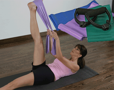

Resistance Band Exercises : Resistance Band Exercises: Teaser with Reverse Fly
[media id=85 width=500 height=400]
Resistance band exercises are widely used by a variety of health and fitness practitioners – both for general strength and conditioning and rehabilitation or injury prevention.
Resistance band exercises are ideal for home exercise programs and can easily be incorporated into a circuit training format helping to condition cardiovascular system as well as strengthening specific muscle groups. Because resistance tubing is so compact and lightweight, it can be used while away from home.
Resistance tubing is extremely adaptable and a large number of resistance band exercises can be developed with very little additional equipment. Smaller muscle groups that are hard to train with more traditional free weight exercises can be targeted with resistance tubing. This makes it particularly appealing to athletic conditioning.
Sports-specific conditioning involves training movements rather than individual muscle groups. The versatility of resistance band exercises allows the athlete to mirror very closely the movement patterns in their sport with varying degrees of resistance. Perhaps even more important is the role they can play in injury prevention and rehabilitation.
Resistance Band Exercises for Injury Prevention & Rehabilitation
Few studies have examined the effects of resistance band exercises on strengthening a specific muscle group. As a result no definite guidelines exist as to the number of sets, repetitions and amount of resistance that should be used. However, many health and fitness practitioners use resistance tubing routinely to prevent and rehabilitate overuse injuries by strengthening often smaller, neglected muscle groups.
For example, athletic movements such as a baseball pitch can place considerable demand on the posterior rotator cuff muscles (external rotators, supraspinatus, infraspinatus, teres minor). These muscles undergo eccentric contraction during the declaration phase of the pitch which places considerable strain on the shoulder . However, the same muscles may not be effectively worked with traditional isotonic exercises . If larger muscle groups, such as the deltoids, become stronger and are able to cope with and apply greater force, this may further compromise the rotator cuff muscles.
A program of resistance band exercises to compliment regular strength training may be able to improve the strength of more isolated muscle groups such as the rotator cuff . Additionally, training these otherwise neglected muscles may even improve performance.
Of course resistance band exercises can be used for more than simply strengthening more isolated muscle groups. The exercises below work the major muscle groups are only a small sample of the many hundreds of variations that have been devised.
Athletes may want to incorporate them into an off season training program when recovery and regeneration is the most important goal.
Resistance bands are available in a range of colors that relate to their stiffness or resistance. Color-coding varies between the brands but it typically as follows:
Yellow (thin)
Red (medium)
Green (heavy)
Blue (extra heavy)
Black (special heavy)
Silver (super heavy)
Band Exercises,Band Exercises Health, Band Exercises Health Latest,Band Exercises Health Information,Band Exercises Health information, Band Exercises Health Photo,Band Exercises for Weight Health photo, Band Exercises Health Latest, Band Exercises Health latest, Band Exercises Video, Band Exercises video, Band Exercises Health History,Band Exercises Health history, Band Exercises over Picture, history, Band Exercises Asia, Band Exercises asia, Band Exercises Gallery, Band Exercises for Weight gallery, Band Exercises Photo Gallery, Band Exercises Picture, Lap picture, Band Exercises Web, Malaysia Health, web Health, web Health picture, video photo, video surgery, gallery, laparoscopy, virus, flu, drug, video, Health Health, calories, photo, nutrition, health video, symptoms, Heart valve, medical, beating, diet, physical, Training, organic, gym, blister, exercise, weightloss, surgery, spiritual, eating, tips, skin, operation,

