Category Archives: Surgery

Surgery

Feet surgery

feet-2

Feet surgery

[media id=25 width=500 height=400]

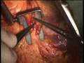
The podiatric surgeon is taught to approach foot surgery by keeping the knowledge of normal foot function and biomechanics in mind. Because of the weightbearing nature of the foot, surgical procedures must be designed to be as stable as possible to withstand the forces of everyday standing and walking. Care is taken to understand the cause of the problem so as to provide a long-lasting cure, when possible. Greater than 99% of podiatric surgery is done in an outpatient setting such as a hospital outpatient department, a freestanding surgery center or in the podiatry office. Most procedures allow for immediate walking with a surgical sandal. Some procedures may require the use of a cane, crutches, or a cast. Specific surgical treatments for many common (and some less common) foot conditions will be discussed.


Flat Feet (Pes Planus)

Surgery for flat feet is generally reserved for the most symptomatic cases. Orthotics are often the first line course of treatment. Many people have what are referred to as “Flat Feet” but are relatively asymptomatic. Flat feet may result in significant foot pain and deformity because of excessive pronation which causes joint instability. Flat foot procedures are designed to provide for a more stable foot which pronates less. Most flat foot surgery is performed on patients in the adolescent age group. There are a large variety of specific surgical procedures that may be used. They may be grouped according to the region of the foot that is treated. Often, 2 or 3 procedures may be performed together from the different groups.

Rearfoot osteotomies
These are procedures which are designed to change the position of the heel into an inverted or supinated position (the opposite of everted and pronated which are found in flat feet.) An osteotomy is a surgical cut in the bone. Often, a wedge of bone is removed to change the angle of the heel bone (calcaneus).  Other procedures are transpositional and involve sliding of one part of the bone along the other part of the bone. (E.g. the Koutsogianis procedure). Other procedures involve adding a bone graft and opening the wedge to change the angle of the calcaneus.( E.g. the Evans Procedure). These osteotomies are generally held together with special screws, pins or bone staples and require a period of casting and immobilization for several weeks.

Medial column stabilizations
These procedures involve fusing two or more of the bones along the medial side (inner side) of the foot. Common fusion sites are the navicular and medial cuneiform.  These bones have often dropped in a flat foot and fusing them provides more stability. These osteotomies are generally held together with special screws, pins or bone staples and require a period of casting and immobilization for several weeks.

Tendon transfers
Sometimes the insertion sites of tendons are detached and then reattached to bones at different locations. The result is a dynamic stabilization. Repositioning of the tendons allows the muscles that pull them to exert their force in a more beneficial way to help support the arch. The Young tenosuspension procedure reattaches the Tibialis Anterior tendon to a better position beneath the medial arch where it can pull up on the arch to support it.

Tendon lengthening
Often, the Achilles tendon is tight and is a major deforming force contributing to flat foot conditions. A condition associated with a tight Achilles tendon is known as equinus. An Achilles tendon lengthening procedure is often effective at reducing this deforming force. The calf is made up of 2 gastrocnemius muscle bellies as well as the soleus muscle. The Achilles tendon attaches to all three. An Achilles tendon lengthening lengthens the whole group together. Sometimes, the gastrocnemius muscles are tight while the soleus is not. In this case, a gastrocnemius recession can be performed to lengthen only the gastrocnemius while leaving the soleus alone.

Arthroeresis
These are procedures in which a peg made of plastic or titanium is placed in front of a bone to limit its motion. A common location for placement of such a device is the Sinus Tarsi which is a cone-shaped space between the talus and calcaneus bones. The peg helps to limit pronation. This is often just a temporary measure with the peg left in for a few years and then removed.

Arthrodeses
An arthrodesis is a fusion of two bones. In addition to the medial column stabilization fusions discussed above, rearfoot bones may also be fused. Rearfoot fusions are generally reserved for the most severely deformed, arthritic and painful feet . A Triple Arthrodesis is a fusion of the Talo-calcaneal, Talo-navicular and Calcaneo-cuboid joints. This is one of the most complex foot surgeries performed since all three joints must be aligned and fused properly to achieve a satisfactory result. In addition, because motion in the rearfoot is eliminated, the ankle joint and other joints in the foot may be forced into compensating to provide additional motion which could result in future symptoms in those places. These fusions are generally held together with special screws, pins or bone staples and require a period of casting and immobilization for two or three months.

Feet surgery , Feet surgery  Health, Feet surgery  Health Latest, Feet surgery  Health Information, Feet surgery  Health information, Feet surgery  Health Photo,Feet surgery  Health photo, Feet surgery  Health Latest, Feet surgery  Health latest, Feet surgery  Health Story, Healthy Minnesota  Health story, Feet surgery  Video, Feet surgery  video, Feet surgery  Health History, Feet surgery  Health history, Feet surgery over Picture, history, Feet surgery  Asia,  Healthy Minnesota  asia, Feet surgery  Gallery, Feet surgery  gallery, Feet surgery  Photo Gallery, Healthy Minnesota  photo gallery, Feet surgery  Picture, Feet surgery  picture, Feet surgery  Web, Malaysia Health, web Health, picture, video photo, video surgery, gallery, laparoscopy, virus, flu, drug, video, Health Health, photo, nutrition, health video, symptoms, cancer, medical, beating, diet, organic, weightloss, surgery, fitness, operation, bf1,  feet,  surgery,  blood,  blister

beating heart surgery

default

beating heart surgery

[media id=24 width=500 height=400]

Beating heart or “off pump” coronary artery surgery is the latest revolution in the management coronary disease. It is being embraced world-wide by increasing numbers of surgeons. Many of the advantages are subtle but reduced mortality, stroke, and bleeding as well as earlier discharge are well-established benefits. A cardiac stabiliser is mandatory for this surgery, most are single use only and very expensive, this one is multiple use and is saving many healthcare dollars.

Beating-heart surgery is a way to perform surgery without stopping the heart. Surgeons use a special device to stabilize the part of the heart on which they are operating. The heart continues to beat and circulate blood to heart muscle during the operation. Surgery on a beating-heart helps reduce the risk for complications associated with temporarily stopping the heart during surgery.

Surgery on a stopped heart is common, and some heart procedures can only be performed on a motionless heart. Physicians use a special solution called cardioplegia to stop the heart.

If the heart is stopped for surgery, the surgeon must restart it and reintroduce blood into the tissue. This is called reperfusion. Reperfusion can cause impairment of heart function. Sometimes, heart muscle tissue can be damaged at the cellular level during reperfusion, a phenomenon known as reperfusion injury. In some people, reperfusion injury can lead to complications such as arrhythmias and heart attacks. Reperfusion injury is especially a concern in high-risk patients, such as the elderly, people who have had several heart surgeries, patients with severe blockages, and those with complex health problems.

Reperfusion injury can be avoided if the heart is kept beating during surgery.

At the University of Chicago Medical Center, our cardiac surgeons opt to perform beating-heart surgery whenever possible. More than 90 percent of coronary artery bypass surgeries performed here are done on a beating heart.

While not all procedures can be performed on a beating heart, our surgeons have developed many techniques that make beating-heart surgery an option for even complex procedures on the inside of the heart–including valve repair. In fact, our surgeons were among the first in the world to perform beating-heart mitral valve surgery. The University of Chicago Medical Center is one of the only hospitals in the nation where beating-heart surgery is being performed to treat valve disease.

Some of the procedures performed on a beating heart include:

Coronary artery bypass graft surgery (including ThoraCAB, a minimally invasive option performed without cutting the breastbone, as well as open-chest, beating-heart bypass)

Surgery for atrial fibrillation

Treatment of some congenital heart defects, such as closure of atrial septal defect

Valve repair (mitral, pulmonary, or tricuspid)

Valve replacement (mitral or tricuspid)

Ventricular reconstruction

Conventional On Pump Coronary Artery Bypass Surgery

More than 70%2 of all bypass surgeries are performed on a stopped heart. Unlike beating heart surgery, during conventional on pump heart bypass, medication is used to stop your heart.

A heart-lung machine takes over the function of your heart and lungs during the surgery.The heart-lung machine is also called a cardiopulmonary bypass machine. It has a pump to function as the heart and a membrane oxygenator to function as the lungs.

Heart-Lung Machine

This mechanical “heart and lungs” keeps oxygen rich blood circulating throughout your body. The heart-lung machine collects the blood. Carbon dioxide and other waste products are removed. The oxygenator adds oxygen, and the oxygenator’s heat exchanger warms (or cools) the blood. The blood is gently circulated back through the body. This process is called perfusion. The person who operates the heart-lung machine is the perfusionist.

Stopping Your Heart

Your heart will usually be stopped for about 30-90 minutes of the 3-6 hour surgery. The heart-lung machine makes it possible for the surgeon to work on a still heart. This technique has been used for many years with excellent results. Once the surgery is over, the surgeon and perfusionist restart your heart.

Medtronic Perfusion Systems

Medtronic creates the dependable perfusion systems that make on pump surgery possible. We are committed to providing doctors, hospitals, and patients with reliable, technically advanced equipment.

beating heart , beating heart  Health, beating heart  Health Latest, beating heart  Health Information, beating heart  Health information, beating heart  Health Photo,beating heart  Health photo, beating heart  Health Latest, beating heart  Health latest, beating heart  Health Story, Healthy Minnesota  Health story, beating heart  Video, beating heart  video, beating heart  Health History, beating heart  Health history, beating heart over Picture, history, beating heart  Asia,  Healthy Minnesota  asia, beating heart  Gallery, beating heart  gallery, beating heart  Photo Gallery, Healthy Minnesota  photo gallery, beating heart  Picture, beating heart  picture, beating heart  Web, Malaysia Health, web Health, picture, video photo, video surgery, gallery, laparoscopy, virus, flu, drug, video, Health Health, photo, nutrition, health video, symptoms, cancer, medical, beating, diet, organic, weightloss, surgery, fitness, operation, bf1, beating,  heart,  surgery,  cabg,  opcab,  bypass

Brain Tumor Surgery

default

Brain Tumor Surgery

[media id=23 width=500 height=400]

A brain tumor is an abnormal growth of cells within the brain or inside the skull, which can be cancerous or non-cancerous (benign).

It is defined as any intracranial tumor created by abnormal and uncontrolled cell division, normally either in the brain itself (neurons, glial cells (astrocytes, oligodendrocytes, ependymal cells), lymphatic tissue, blood vessels), in the cranial nerves (myelin-producing Schwann cells), in the brain envelopes (meninges), skull, pituitary and pineal gland, or spread from cancers primarily located in other organs (metastatic tumors).

Primary (true) brain tumors are commonly located in the posterior cranial fossa in children and in the anterior two-thirds of the cerebral hemispheres in adults, although they can affect any part of the brain.

Signs and symptoms

Symptoms of brain tumors may depend on two factors: tumor size (volume) and tumor location. The time point of symptom onset in the course of disease correlates in many cases with the nature of the tumor (“benign”, i.e. slow-growing/late symptom onset, or malignant, fast growing/early symptom onset) is a frequent reason for seeking medical attention in brain tumor cases.

Large tumors or tumors with extensive perifocal swelling edema inevitably lead to elevated intracranial pressure (intracranial hypertension), which translates clinically into headaches, vomiting (sometimes without nausea), altered state of consciousness (somnolence, coma), dilatation of the pupil on the side of the lesion (anisocoria), papilledema (prominent optic disc at the funduscopic examination). However, even small tumors obstructing the passage of cerebrospinal fluid (CSF) may cause early signs of increased intracranial pressure. Increased intracranial pressure may result in herniation (i.e. displacement) of certain parts of the brain, such as the cerebellar tonsils or the temporal uncus, resulting in lethal brainstem compression. In young children, elevated intracranial pressure may cause an increase in the diameter of the skull and bulging of the fontanelles.

A bilateral temporal visual field defect (bitemporal hemianopia—due to compression of the optic chiasm), often associated with endocrine disfunction—either hypopituitarism or hyperproduction of pituitary hormones and hyperprolactinemia is suggestive of a pituitary tumor.

Treatment and prognosis

Many meningiomas, with the exception of some tumors located at the skull base, can be successfully removed surgically. In more difficult cases, stereotactic radiosurgery, such as Gamma knife, Cyberknife or Novalis Tx radiosurgery, remains a viable option.

Most pituitary adenomas can be removed surgically, often using a minimally invasive approach through the nasal cavity and skull base (trans-nasal, trans-sphenoidal approach). Large pituitary adenomas require a craniotomy (opening of the skull) for their removal. Radiotherapy, including stereotactic approaches, is reserved for the inoperable cases.

Brain Tumor , Brain Tumor  Health, Brain Tumor  Health Latest, Brain Tumor  Health Information, Brain Tumor  Health information, Brain Tumor  Health Photo,Brain Tumor  Health photo, Brain Tumor  Health Latest, Brain Tumor  Health latest, Brain Tumor  Health Story, Healthy Minnesota  Health story, Brain Tumor  Video, Brain Tumor  video, Brain Tumor  Health History, Brain Tumor  Health history, Brain Tumor over Picture, history, Brain Tumor  Asia,  Healthy Minnesota  asia, Brain Tumor  Gallery, Brain Tumor  gallery, Brain Tumor  Photo Gallery, Healthy Minnesota  photo gallery, Brain Tumor  Picture, Brain Tumor  picture, Brain Tumor  Web, Malaysia Health, web Health, picture, video photo, video surgery, gallery, laparoscopy, virus, flu, drug, video, Health Health, photo, nutrition, health video, symptoms, cancer, medical, diet, organic, weightloss, surgery, fitness, operation, bf1, Kennedy,  brain,  tumor,  Ted,  Brain,  Cancer,  Malignan,  cancer,  Biopsy , glioma