Tag Archives: Brain

Beyond the Brain

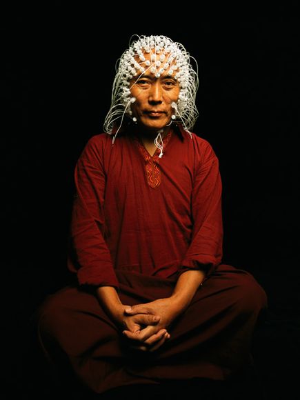
Electrodes measure a Tibetan monk’s brain activity.

The ancient Egyptians thought so little of brain matter they made a practice of scooping it out through the nose of a dead leader before packing the skull with cloth before burial. They believed consciousness resided in the heart, a view shared by Aristotle and a legacy of medieval thinkers. Even when consensus for the locus of thought moved northward into the head, it was not the brain that was believed to be the sine qua non, but the empty spaces within it, called ventricles, where ephemeral spirits swirled about. As late as 1662, philosopher Henry More scoffed that the brain showed “no more capacity for thought than a cake of suet, or a bowl of curds.”

Around the same time, French philosopher René Descartes codified the separation of conscious thought from the physical flesh of the brain. Cartesian “dualism” exerted a powerful influence over Western science for centuries, and while dismissed by most neuroscientists today, still feeds the popular belief in mind as a magical, transcendent quality.

A contemporary of Descartes named Thomas Willis—often referred to as the father of neurology—was the first to suggest that not only was the brain itself the locus of the mind, but that different parts of the brain give rise to specific cognitive functions. Early 19th-century phrenologists pushed this notion in a quaint direction, proposing that personality proclivities could be deduced by feeling the bumps on a person’s skull, which were caused by the brain “pushing out” in places where it was particularly well developed. Plaster casts of the heads of executed criminals were examined and compared to a reference head to determine whether any particular protuberances could be reliably associated with criminal behavior.

Though absurdly unscientific even for its time, phrenology was remarkably prescient—up to a point. In the past decade especially, advanced technologies for capturing a snapshot of the brain in action have confirmed that discrete functions occur in specific locations. The neural “address” where you remember a phone number, for instance, is different from the one where you remember a face, and recalling a famous face involves different circuits than remembering your best friend’s.

Yet it is increasingly clear that cognitive functions cannot be pinned to spots on the brain like towns on a map. A given mental task may involve a complicated web of circuits, which interact in varying degrees with others throughout the brain—not like the parts in a machine, but like the instruments in a symphony orchestra combining their tenor, volume, and resonance to create a particular musical effect.

Corina’s brain all she is…is here

Corina Alamillo is lying on her right side in an operating room in the UCLA Medical Center. There is a pillow tucked beneath her cheek and a steel scaffold screwed into her forehead to keep her head perfectly still. A medical assistant in her late 20s, she has dark brown eyes, full eyebrows, and a round, open face.

Brain Tumor Surgery

default

Brain Tumor Surgery

[media id=23 width=500 height=400]

A brain tumor is an abnormal growth of cells within the brain or inside the skull, which can be cancerous or non-cancerous (benign).

It is defined as any intracranial tumor created by abnormal and uncontrolled cell division, normally either in the brain itself (neurons, glial cells (astrocytes, oligodendrocytes, ependymal cells), lymphatic tissue, blood vessels), in the cranial nerves (myelin-producing Schwann cells), in the brain envelopes (meninges), skull, pituitary and pineal gland, or spread from cancers primarily located in other organs (metastatic tumors).

Primary (true) brain tumors are commonly located in the posterior cranial fossa in children and in the anterior two-thirds of the cerebral hemispheres in adults, although they can affect any part of the brain.

Signs and symptoms

Symptoms of brain tumors may depend on two factors: tumor size (volume) and tumor location. The time point of symptom onset in the course of disease correlates in many cases with the nature of the tumor (“benign”, i.e. slow-growing/late symptom onset, or malignant, fast growing/early symptom onset) is a frequent reason for seeking medical attention in brain tumor cases.

Large tumors or tumors with extensive perifocal swelling edema inevitably lead to elevated intracranial pressure (intracranial hypertension), which translates clinically into headaches, vomiting (sometimes without nausea), altered state of consciousness (somnolence, coma), dilatation of the pupil on the side of the lesion (anisocoria), papilledema (prominent optic disc at the funduscopic examination). However, even small tumors obstructing the passage of cerebrospinal fluid (CSF) may cause early signs of increased intracranial pressure. Increased intracranial pressure may result in herniation (i.e. displacement) of certain parts of the brain, such as the cerebellar tonsils or the temporal uncus, resulting in lethal brainstem compression. In young children, elevated intracranial pressure may cause an increase in the diameter of the skull and bulging of the fontanelles.

A bilateral temporal visual field defect (bitemporal hemianopia—due to compression of the optic chiasm), often associated with endocrine disfunction—either hypopituitarism or hyperproduction of pituitary hormones and hyperprolactinemia is suggestive of a pituitary tumor.

Treatment and prognosis

Many meningiomas, with the exception of some tumors located at the skull base, can be successfully removed surgically. In more difficult cases, stereotactic radiosurgery, such as Gamma knife, Cyberknife or Novalis Tx radiosurgery, remains a viable option.

Most pituitary adenomas can be removed surgically, often using a minimally invasive approach through the nasal cavity and skull base (trans-nasal, trans-sphenoidal approach). Large pituitary adenomas require a craniotomy (opening of the skull) for their removal. Radiotherapy, including stereotactic approaches, is reserved for the inoperable cases.

Brain Tumor , Brain Tumor  Health, Brain Tumor  Health Latest, Brain Tumor  Health Information, Brain Tumor  Health information, Brain Tumor  Health Photo,Brain Tumor  Health photo, Brain Tumor  Health Latest, Brain Tumor  Health latest, Brain Tumor  Health Story, Healthy Minnesota  Health story, Brain Tumor  Video, Brain Tumor  video, Brain Tumor  Health History, Brain Tumor  Health history, Brain Tumor over Picture, history, Brain Tumor  Asia,  Healthy Minnesota  asia, Brain Tumor  Gallery, Brain Tumor  gallery, Brain Tumor  Photo Gallery, Healthy Minnesota  photo gallery, Brain Tumor  Picture, Brain Tumor  picture, Brain Tumor  Web, Malaysia Health, web Health, picture, video photo, video surgery, gallery, laparoscopy, virus, flu, drug, video, Health Health, photo, nutrition, health video, symptoms, cancer, medical, diet, organic, weightloss, surgery, fitness, operation, bf1, Kennedy,  brain,  tumor,  Ted,  Brain,  Cancer,  Malignan,  cancer,  Biopsy , glioma

Brain Stem Tumor Operation

images

Brain Stem Tumor Operation

[media id=13 width=500 height=400]

Brain Stem Tumor Operation in sitting position by Prof Nazir Ahmad Lahore Pakistan.

Brain tumors are tumors that grow in the brain. A tumor is an abnormal growth caused by cells reproducing themselves in an uncontrolled manner.

When doctors describe tumors in parts of the body other than the brain, they use the words benign (meaning harmless) or malignant (meaning cancerous). These meanings change, however, when referring to tumors in the brain.

Benign brain tumors:

A benign brain tumor consists of benign (harmless) cells and has distinct boundaries. Surgery alone may cure this type of tumor.

Malignant brain tumors:

A malignant brain tumor is life-threatening. It may be malignant because it consists of cancer cells, or it may be called malignant because of its location. In other words, a brain tumor composed of benign cells–but located in a vital area–is still considered malignant.

A malignant brain tumor made up of cancerous cells may spread or seed (metastasize) to other locations in the brain or spinal cord. It can invade and destroy healthy tissue so it cannot function properly. Malignant tumors grow the way a plant does, with “roots” invading various tissues. Or, they can shed cells that travel to distant parts of the brain. Some cancerous tumors, however, remain localized.

Brain Stem Tumor, Brain Stem Tumor Health, Brain Stem Tumor Health Latest, Brain Stem Tumor Health Information, bealthy breakfast Health information, Brain Stem Tumor Health Photo, Brain Stem Tumor Health Latest, Brain Stem Tumor Health latest, Brain Stem Tumor Health Story, Brain Stem Tumor Health story, Brain Stem Tumor Video, Brain Stem Tumor video, Brain Stem Tumor Health History, Brain Stem Tumor Health history, history, Brain Stem Tumor Asia,  Brain Stem Tumor asia, Brain Stem Tumor Gallery, Brain Stem Tumor gallery, Brain Stem Tumor Photo Gallery, Brain Stem Tumor photo gallery, Brain Stem Tumor Picture, Brain Stem Tumor picture, Brain Stem Tumor Web, Malaysia Health, web Health, picture, video photo, gallery, virus, flu, drug, symptoms , video, photo, surgery, operation, bf1, health , Neurosurgery,  Brain,  Stem,  Tumor,  Operation