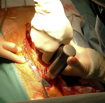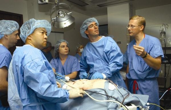

Lung Surgery Operation
[media id=39 width=500 height=400]
Lung
The lung or pulmonary system is the essential respiration organ in air-breathing animals, including most tetrapods, a few fish and a few snails. In mammals and the more complex life forms, the two lungs are located in the chest on either side of the heart. Their principal function is to transport oxygen from the atmosphere into the bloodstream, and to release carbon dioxide from the bloodstream into the atmosphere. This exchange of gases is accomplished in the mosaic of specialized cells that form millions of tiny, exceptionally thin-walled air sacs called alveoli.
In order to completely explain the anatomy of the lungs, it is necessary to discuss the passage of air through the mouth to the alveoli. Once air progresses through the mouth or nose, it travels through the oropharynx, nasopharynx, the larynx, the trachea, and a progressively subdividing system of bronchi and bronchioles until it finally reaches the alveoli where the gas exchange of carbon dioxide and oxygen takes place.]
The drawing and expulsion of air (ventilation) is driven by muscular action; in early tetrapods, air was driven into the lungs by the pharyngeal muscles, whereas in reptiles, birds and mammals a more complicated musculoskeletal system is used.
Medical terms related to the lung often begin with pulmo-, from the Latin pulmonarius (“of the lungs”), or with pneumo- (from Greek πνεύμων “lung”).
Anatomy
In humans, the trachea divides into the two main bronchi that enter the roots of the lungs. The bronchi continue to divide within the lung, and after multiple divisions, give rise to bronchioles. The bronchial tree continues branching until it reaches the level of terminal bronchioles, which lead to alveolar sacs. Alveolar sacs are made up of clusters of alveoli, like individual grapes within a bunch. The individual alveoli are tightly wrapped in blood vessels and it is here that gas exchange actually occurs. Deoxygenated blood from the heart is pumped through the pulmonary artery to the lungs, where oxygen diffuses into blood and is exchanged for carbon dioxide in the hemoglobin of the erythrocytes. The oxygen-rich blood returns to the heart via the pulmonary veins to be pumped back into systemic circulation.
Human lungs are located in two cavities on either side of the heart. Though similar in appearance, the two are not identical. Both are separated into lobes by fissures, with three lobes on the right and two on the left. The lobes are further divided into segments and then into lobules, hexagonal divisions of the lungs that are the smallest subdivision visible to the naked eye. The connective tissue that divides lobules is often blackened in smokers and city dwellers. The medial border of the right lung is nearly vertical, while the left lung contains a cardiac notch. The cardiac notch is a concave impression molded to accommodate the shape of the heart. Lungs are to a certain extent ‘overbuilt’ and have a tremendous reserve volume as compared to the oxygen exchange requirements when at rest. Such excess capacity is one of the reasons that individuals can smoke for years without having a noticeable decrease in lung function while still or moving slowly; in situations like these only a small portion of the lungs are actually perfused with blood for gas exchange. As oxygen requirements increase due to exercise, a greater volume of the lungs is perfused, allowing the body to match its CO2/O2 exchange requirements.
The environment of the lung is very moist, which makes it hospitable for bacteria. Many respiratory illnesses are the result of bacterial or viral infection of the lungs. Inflammation of the lungs is known as pneumonia; inflammation of the pleura surrounding the lungs is known as pleurisy.
Vital capacity is the maximum volume of air that a person can exhale after maximum inhalation; it can be measured with a spirometer. In combination with other physiological measurements, the vital capacity can help make a diagnosis of underlying lung disease.
Lung Surgery , Lung Surgery Health, Lung Surgery Health Latest, Lung Surgery Health Information, Lung Surgery Health information, Lung Surgery Health Photo,Exercising for Weight Health photo, Lung Surgery Health Latest, Lung Surgery Health latest, Exercising for Weight Health Story, Healthy Minnesota Health story, Lung Surgery Video, Lung Surgery video, Lung Surgery Health History, Lung Surgery Health history, Lung Surgery over Picture, history, Lung Surgery Asia, Healthy Minnesota asia, Lung Surgery Gallery, Exercising for Weight gallery, Lung Surgery Photo Gallery, Healthy Minnesota photo gallery, Lung Surgery Picture, Lung Surgery picture, Lung Surgery Web, Malaysia Health, web Health, web Health picture, video photo, video surgery, gallery, laparoscopy, virus, flu, drug, video, Health Health, calories, photo, nutrition, health video, symptoms, cancer, medical, beating, diet, physical, Training, organic, gym, blister, exercise, weightloss, surgery, spiritual, eating, tips, skin, operation, bf1, Lung, Surgery , Operation , Inside, Body