Category Archives: Surgery

Surgery

Lung Surgery Operation

070629193359-large

Lung Surgery Operation

[media id=39 width=500 height=400]

Lung

The lung or pulmonary system is the essential respiration organ in air-breathing animals, including most tetrapods, a few fish and a few snails. In mammals and the more complex life forms, the two lungs are located in the chest on either side of the heart. Their principal function is to transport oxygen from the atmosphere into the bloodstream, and to release carbon dioxide from the bloodstream into the atmosphere. This exchange of gases is accomplished in the mosaic of specialized cells that form millions of tiny, exceptionally thin-walled air sacs called alveoli.

In order to completely explain the anatomy of the lungs, it is necessary to discuss the passage of air through the mouth to the alveoli. Once air progresses through the mouth or nose, it travels through the oropharynx, nasopharynx, the larynx, the trachea, and a progressively subdividing system of bronchi and bronchioles until it finally reaches the alveoli where the gas exchange of carbon dioxide and oxygen takes place.]

The drawing and expulsion of air (ventilation) is driven by muscular action; in early tetrapods, air was driven into the lungs by the pharyngeal muscles, whereas in reptiles, birds and mammals a more complicated musculoskeletal system is used.

Medical terms related to the lung often begin with pulmo-, from the Latin pulmonarius (“of the lungs”), or with pneumo- (from Greek πνεύμων “lung”).

Anatomy

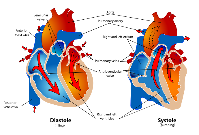
In humans, the trachea divides into the two main bronchi that enter the roots of the lungs. The bronchi continue to divide within the lung, and after multiple divisions, give rise to bronchioles. The bronchial tree continues branching until it reaches the level of terminal bronchioles, which lead to alveolar sacs. Alveolar sacs are made up of clusters of alveoli, like individual grapes within a bunch. The individual alveoli are tightly wrapped in blood vessels and it is here that gas exchange actually occurs. Deoxygenated blood from the heart is pumped through the pulmonary artery to the lungs, where oxygen diffuses into blood and is exchanged for carbon dioxide in the hemoglobin of the erythrocytes. The oxygen-rich blood returns to the heart via the pulmonary veins to be pumped back into systemic circulation.

Human lungs are located in two cavities on either side of the heart. Though similar in appearance, the two are not identical. Both are separated into lobes by fissures, with three lobes on the right and two on the left. The lobes are further divided into segments and then into lobules, hexagonal divisions of the lungs that are the smallest subdivision visible to the naked eye. The connective tissue that divides lobules is often blackened in smokers and city dwellers. The medial border of the right lung is nearly vertical, while the left lung contains a cardiac notch. The cardiac notch is a concave impression molded to accommodate the shape of the heart. Lungs are to a certain extent ‘overbuilt’ and have a tremendous reserve volume as compared to the oxygen exchange requirements when at rest. Such excess capacity is one of the reasons that individuals can smoke for years without having a noticeable decrease in lung function while still or moving slowly; in situations like these only a small portion of the lungs are actually perfused with blood for gas exchange. As oxygen requirements increase due to exercise, a greater volume of the lungs is perfused, allowing the body to match its CO2/O2 exchange requirements.

The environment of the lung is very moist, which makes it hospitable for bacteria. Many respiratory illnesses are the result of bacterial or viral infection of the lungs. Inflammation of the lungs is known as pneumonia; inflammation of the pleura surrounding the lungs is known as pleurisy.

Vital capacity is the maximum volume of air that a person can exhale after maximum inhalation; it can be measured with a spirometer. In combination with other physiological measurements, the vital capacity can help make a diagnosis of underlying lung disease.

Lung Surgery  , Lung Surgery   Health, Lung Surgery   Health Latest, Lung Surgery   Health Information, Lung Surgery   Health information, Lung Surgery  Health Photo,Exercising for Weight Health photo, Lung Surgery   Health Latest, Lung Surgery  Health latest, Exercising for Weight  Health Story, Healthy Minnesota  Health story, Lung Surgery   Video, Lung Surgery   video, Lung Surgery   Health History, Lung Surgery   Health history, Lung Surgery  over Picture, history, Lung Surgery   Asia,  Healthy Minnesota  asia, Lung Surgery   Gallery, Exercising for Weight  gallery, Lung Surgery   Photo Gallery, Healthy Minnesota  photo gallery, Lung Surgery   Picture, Lung Surgery   picture, Lung Surgery   Web, Malaysia Health, web Health, web Health picture, video photo, video surgery, gallery, laparoscopy, virus, flu, drug, video, Health Health, calories, photo, nutrition, health video, symptoms, cancer, medical, beating, diet, physical, Training, organic, gym, blister, exercise, weightloss, surgery, spiritual, eating, tips, skin, operation, bf1, Lung,  Surgery , Operation , Inside,  Body

Median Sternotomy

sternotomy

Median Sternotomy

[media id=37 width=500 height=400]

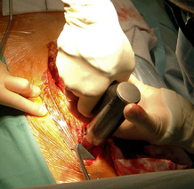
Median sternotomy is a type of surgical procedure in which a vertical inline incision is made along the sternum, after which the sternum itself is divided, or “cracked”. This procedure provides access to the heart and lungs for surgical procedures such as heart transplant, corrective surgery for congenital heart defects (CHDs), or coronary artery bypass surgery.

Median sternotomy is often mistakenly referred to as open heart surgery; however, open heart involves incision of the pericardium, and many median sternotomy procedures do not require this. Open heart usually involves the use of a cardiopulmonary bypass, also known as a heart-lung machine.

Median Sternotomy , Median Sternotomy  Health, Median Sternotomy  Health Latest, Median Sternotomy  Health Information, Median Sternotomy  Health information, Median Sternotomy Health Photo,Exercising for Weight Health photo, Median Sternotomy  Health Latest, Median Sternotomy Health latest, Exercising for Weight  Health Story, Healthy Minnesota  Health story, Median Sternotomy  Video, Median Sternotomy  video, Median Sternotomy  Health History, Median Sternotomy  Health history, Median Sternotomy over Picture, history, Median Sternotomy  Asia,  Healthy Minnesota  asia, Median Sternotomy  Gallery, Exercising for Weight  gallery, Median Sternotomy  Photo Gallery, Healthy Minnesota  photo gallery, Median Sternotomy  Picture, Median Sternotomy  picture, Median Sternotomy  Web, Malaysia Health, web Health, picture, video photo, video surgery, gallery, laparoscopy, virus, flu, drug, video, Health Health, calories, photo, nutrition, health video, symptoms, cancer, medical, beating, diet, organic, blister, exercise, weightloss, surgery, spiritual, eating, tips, skin, operation, bf1, Sternotomy

Pumping Heart

Human_healthy_pumping_heart

Pumping Heart

[media id=36 width=500 height=400]

A pump is a device used to move fluids, such as gases, liquids or slurries. A pump displaces a volume by physical or mechanical action. One common misconception about pumps is the thought that they create pressure. Pumps alone do not create pressure; they only displace fluid, causing a flow. Adding resistance to flow causes pressure. Pumps fall into two major groups: positive displacement pumps and rotodynamic pumps. Their names describe the method for moving a fluid.

A positive displacement pump causes a fluid to move by trapping a fixed amount of it then forcing (displacing) that trapped volume into the discharge pipe. A positive displacement pump can be further classified according to the mechanism used to move the fluid:

Rotary-type, for example, the lobe, external gear, internal gear, screw, shuttle block, flexible vane or sliding vane, helical twisted roots (e.g. the Wendelkolben pump) or liquid ring vacuum pumps.

Positive displacement rotary pumps are pumps that move fluid using the principles of rotation. The vacuum created by the rotation of the pump captures and draws in the liquid. Rotary pumps are very efficient because they naturally remove air from the lines, eliminating the need to bleed the air from the lines manually. Positive displacement rotary pumps also have their weaknesses. Because of the nature of the pump, the clearance between the rotating pump and the outer edge must be very close, requiring that the pumps rotate at a slow, steady speed. If rotary pumps are operated at high speeds, the fluids will cause erosion, much as ocean waves polish stones or erode rock into sand. Rotary pumps that experience such erosion eventually show signs of enlarged clearances, which allow liquid to slip through and detract from the efficiency of the pump. Positive displacement rotary pumps can be grouped into three main types. Gear pumps are the simplest type of rotary pumps, consisting of two gears laid out side-by-side with their teeth enmeshed.

The gears turn away from each other, creating a current that traps fluid between the teeth on the gears and the outer casing, eventually releasing the fluid on the discharge side of the pump as the teeth mesh and go around again. Many small teeth maintain a constant flow of fluid, while fewer, larger teeth create a tendency for the pump to discharge fluids in short, pulsing gushes. Screw pumps are a more complicated type of rotary pumps, featuring two screws with opposing thread —- that is, one screw turns clockwise, and the other counterclockwise. The screws are each mounted on shafts that run parallel to each other; the shafts also have gears on them that mesh with each other in order to turn the shafts together and keep everything in place. The turning of the screws, and consequently the shafts to which they are mounted, draws the fluid through the pump. As with other forms of rotary pumps, the clearance between moving parts and the pump’s casing is minimal. Moving vane pumps are the third type of rotary pumps, consisting of a cylindrical rotor encased in a similarly shaped housing. As the rotor turns, the vanes trap fluid between the rotor and the casing, drawing the fluid through the pump.

Pumping Heart , Pumping Heart  Health, Pumping Heart  Health Latest, Pumping Heart  Health Information, Pumping Heart  Health information, Pumping Heart Health Photo,Exercising for Weight Health photo, Pumping Heart  Health Latest, Pumping Heart Health latest, Exercising for Weight  Health Story, Healthy Minnesota  Health story, Pumping Heart  Video, Pumping Heart  video, Pumping Heart  Health History, Pumping Heart  Health history, Pumping Heart over Picture, history, Pumping Heart  Asia,  Healthy Minnesota  asia, Pumping Heart  Gallery, Exercising for Weight  gallery, Pumping Heart  Photo Gallery, Healthy Minnesota  photo gallery, Pumping Heart  Picture, Pumping Heart  picture, Pumping Heart  Web, Malaysia Health, web Health, picture, video photo, video surgery, gallery, laparoscopy, virus, flu, drug, video, Health Health, calories, photo, nutrition, health video, symptoms, cancer, medical, beating, diet, organic, blister, exercise, weightloss, surgery, spiritual, eating, tips, skin, operation, bf1,heart